
Prior to clinical neuroscience
The importance of MRI technology in preclinical research in neuroscience
Neuroscience research how to help us learn more about brain function
We can use magnetic resonance imaging (MRI) to provide 2 d or 3 d image of the brain, molecular mechanism is used to study the anatomical structure, function, or...Or a combination of all three.The benefits of MRI is that researchers can choose to focus on the anatomical and functional level and molecular level.
In vivo neuroimaging can provide us with what information about the function and metabolism of the brain?
Using a technique called diffusion MRI, we can be in the form of non invasive and non destructive, tracking the whole brain axons direction, and create the connection diagram of the brain.
In terms of functional, we have many choices.Functional MRI (fMRI) allows us to observe it in the brain to think.The technology belongs to the clinical criteria, in the past ten years, we've been able to applied to animals, including rats and mice.FMRI don't need a contrast agent.We need to monitor because of oxyhemoglobin and deaeration hemoglobin transformation and the small signal change, can clearly detect brain activity.
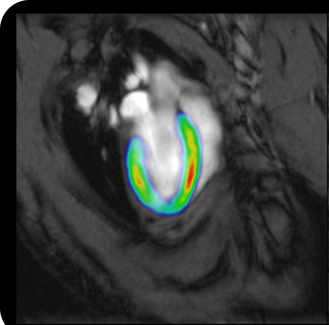
In addition, we also can monitor the change of cerebral blood flow, it is an important symbol.In stroke study, we can see that the affected areas of the brain, its precision is higher than in most other non-destructive method.
Living spectrum to study the metabolites in the body.Take this, we can obtain the area of the brain chemical "fingerprint".These areas are usually a few cubic millimeter, the size of the local living spectrum allows us to identify and quantify the dozens of metabolites, including mainly related to the energy pathways of the brain neurotransmitter and molecules.
Not all biologists to understand MRI technology, why?
Magnetic resonance imaging (MRI) usually do not belong to the category of biology course.M.d., Ph.D., will receive basic training on MRI, if finally become a radiologist, will undergo further training.But for biologists, they contact with MRI in be used to solve the problem of biology.I used to and biology and chemistry courses, and all the basic knowledge about NMR and MRI, I am in chemistry lessons learned.If I only learn biology, I don't know anything about the great potential of MRI.
Each biologists will learn how to use optical microscope, but unless equipped with before clinical MRI scanner located in university, they can hardly to understand MRI techniques.Brooke MRI applications experts have put their knowledge into the optimization of the agreement in advance, even if the user not understanding of MRI, also can quickly solve biology related issues.
Please summarize MRI and PET/MRI application and importance in basic neuroscience research.
PET information lack of anatomy.In general, the use of PET, you can track tracer in any location in the body, and you finally see is functional tracer area.
If you use PET alone, cannot determine the active area in the body position, because there is no anatomy related references.And, using a combination of PET/MRI in gray coloured PET images of high-resolution MRI images, you can see the location of the tracer accurately.
PET and MRI combined with the importance and wonderful is that you can perform these two operations at the same time, and obtain good soft tissue from MRI contrast.
Compared with other methods, these imaging techniques have what advantage?
Besides non destructive, and the fact is that we can use fewer animals for more information.
You can achieve greater statistical correlation, because you can use the scanner in weeks or months study repeatedly the same animal.After each research point, animals will not be disposal.On the contrary, we scan the entire queue, get all information from all the animals there.Every animal as their own controls.This reduces the many pre-clinical inherent biological scattering problems.I think this is an often overlooked the huge advantage.
Preclinical research findings can completely into clinical application?Clinical forebrain imaging can be clinically impossible?
Yes, can be changed.For animal use PET and MRI images and in the hospital to the operation of the patients with clinical instruments were the same.Preclinical imaging, of course, also have advantages, such as before entering the clinical test new methods of disease treatment.You can also use the knockout model to study the mechanism of disease progression.
Please introduce for before clinical neuroscience research brooke instruments:
As early as more than 40 years ago, we launched a series of preclinical MRI scanner, a leading position in the market.Brooke's clinical MRI scanners before brand called BioSpecs, there are different versions.You can choose from a series of magnetic field.The stronger the magnetic field, usually imaging the better the results.You also need to determine the pore diameter, that is, a magnet inside the small channel, animals in the inspection when it's lying in it.Small aperture scanner can only hold a mouse, and other large aperture scanner can accommodate rats and even larger animals.
Our PET scanner also has small channels for the use of rats and mice.We also offer a combination of PET and MRI.In one design of PET/MR, PET channel in MRI in the front of the channel, so the two machines are adjacent to each other.Animals in a similar orbit of monorail, first enter the quick scan of the PET passage.Then move forward about 20 inches, positioning in the MRI scanner, performed MRI scans.
In another PET/MR design, small PET ring directly installed in MRI channels, make animals can directly into the center of the MRI scanner, which is the center of the PET scanner, can realize scanning at the same time.
This instrument has been used to study the preclinical disease model?Whether can help identify any potential treatment?
Well, the application is very broad, from alzheimer's disease and Parkinson's disease model to memory, aging, and cognitive decline model, and so on.The instrument is used in the study of stroke.By artificially induced stroke in rodents, we can to quantify the affected areas of the brain, this perhaps more than any other method that does not involve the anatomy of the brain are more efficient.Many pharmaceutical companies use brooke scanner in drug development.

LabScape
Brooke BioSpin nuclear magnetic resonance (NMR) and preclinical imaging products, services and lifecycle support
Brooke promises to provide clients with unparalleled throughout the buying cycle, from the initial consultation to the evaluation, installation, and service life of the instrument, it is LabScape always adhere to the service concept.
LabScape Maintenance agreement (Maintenance Agreements), the optional services (On - site On - Demand) and laboratory improvement plan (Enhance Your Lab) modern Lab is dedicated to provide you with a new method of Maintenance and service.