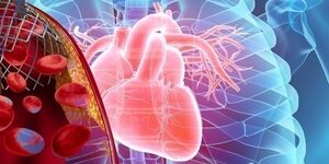
Have a better understanding of cardiovascular disease
In 2020, the cardiovascular disease killed more than 19 million people worldwide1
Before the clinical cardiovascular research
In 2020, cardiovascular disease (CVD) killed about 19.1 million people worldwide1According to the world health organization (WHO), data, cardiovascular disease is the world's leading cause of death.2The high morbidity and high mortality in low and middle income countries is on the rise, the population of the country lived significantly longer than in the past, therefore, are at higher risk of cardiovascular disease.
However, age is not the only risk factor.Causes coronary heart disease and cerebrovascular disease such as the root cause of the different types of cardiovascular disease can be very complex, and not just eating and smoking.It turned out that poverty, stress and pollution of the environment are all factors affecting the risk of cardiovascular disease.
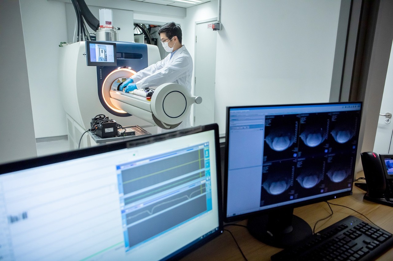
Magnetic resonance imaging (MRI) is a widely used in cardiovascular disease diagnosis method, also is the important tool of preclinical studies.By using resonance spectroscopy (MRS) and magnetic resonance angiography (MRA), such as magnetic resonance imaging technology research with cardiovascular disease animal model, researchers can better understand the pathophysiology of diseases and the progress of the disease.
This non-invasive method can be in a long time, the longitudinal measurement, and provide a large number of quantifiable parameters, such as, ventricular volume or strain.
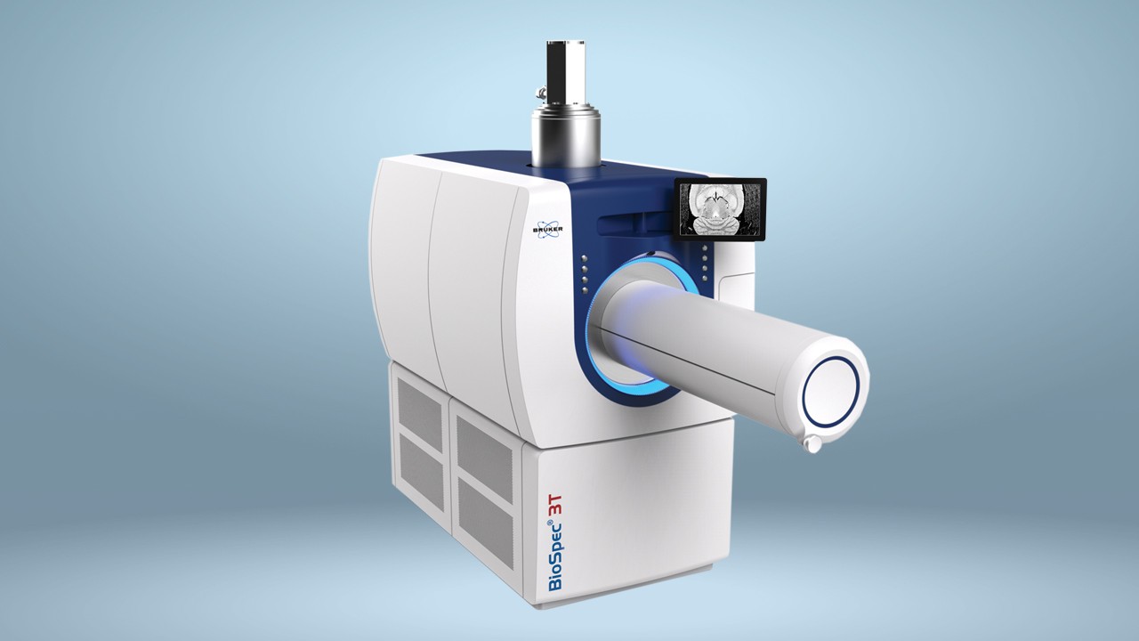
A new generation of MR technology
Preclinical MR imaging is a mature technology, used to study a variety of small animal model of heart disease.Although usually in clinical CT electrocardiogram (ecg) scheme using CT contrast agent (for example, the iodine contrast agent) for clean up too fast, does not apply to small animal models, but before the clinical MR for contrast enhanced imaging and a contrast enhancement imaging, provides powerful heart.
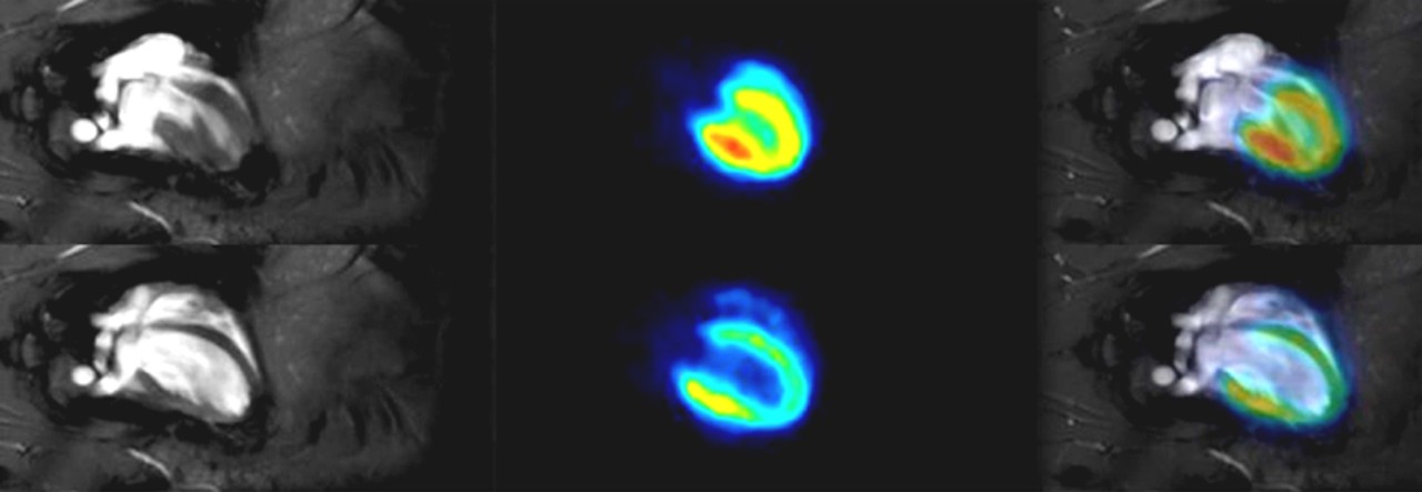
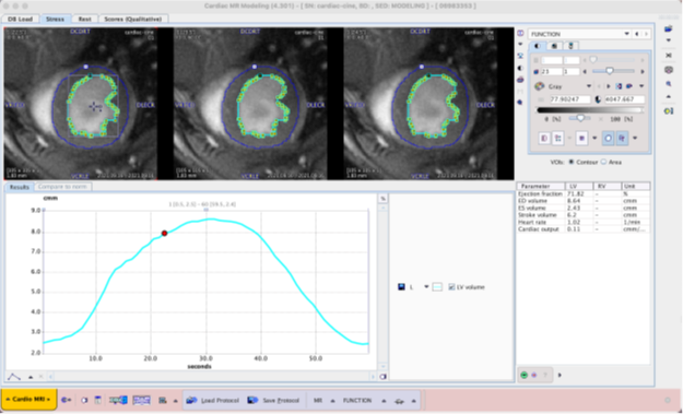
Because of mice and rats heart rate five times faster than humans, therefore, often need to use a good slice and/or frame coverage of high contrast CINE imaging, to parse the rodents microscopic characteristics of cardiac tissue, the function such as ejection fraction measurement.

Compared with the traditional CINE imaging (yellow), CINE imaging (red) can accelerate the SMS in the absence of prolonged scanning, provide greater slice coverage.Scanning through the BioSpec 70/30 array, and the hearts of rats changed only SMS factor (no) or SMS2 and section number (2 or 4).All other parameters are constant, TE/TR: 1.6/10 ms, resolution (130 x 130) mu m2, slice thickness: 800 microns;Film frames: 12, acquisition time (trigger) : about 8 m 30 s.
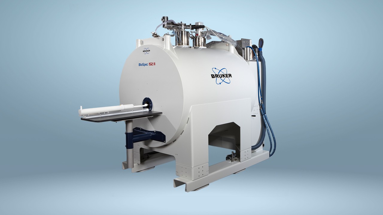
The working process for the MR
MR before clinical method of hardware and software implementation on maximum shortening the total scan time at the same time, achieving high signal-to-noise ratio (SNR), it will be great help for small animal cardiology.Brooke's MR instrument hardware and methods, including the heart of the accord with human body engineering design, rapid gradient array coil and amplifier, since the door control method (namely IntraGate) and segmentation based on artificial intelligence (AI), can significantly shorten the total measurement time, improve cardiac imaging performance.
In addition to clinical heart before measuring the improvement of the method itself, from setting to cardiac evaluation parameters, the compact and powerful workflow, make heart study and analysis of a simple process, provides support for preclinical research personnel.
reference
- 2022 Heart diseases and Stroke Statistical Update Fact Sheet: Global Burden of diseases, the American Heart Association.https://www.heart.org/-/media/PHD-Files-2/Science-雷竞技官方网站News/2/2022-Heart-and-Stroke-Stat-Update/2022-Stat-Update-factsheet-GIobal-Burden-of-Disease.pdf[Accessed 11.02.22]
- Cardiovascular diseases (CVDs) : the Key facts. The World Health Organization (WHO), June 2021.https://www.who.int/雷竞技官方网站news-room/fact-sheets/detail/cardiovascular-diseases- (CVDS)[Accessed 11.02.22]