ELIO
The only element of portable XRF imaging spectrometer on the market
Element is used to analyze the quality of the spectrometer

Bright spot
With the imaging features of portable XRF spectrometer faded area

The key characteristics of
- Truly portable and flexible
- Non-contact measurement
- nondestructive
- No sample preparation
- The laser focus and microscopic camera, is used for precise control
- A sum of element distribution and the optical image
- Reliable detection of trace element
Brooke ELIO easy to operate, and can reveal the important information of the objects you are testing.When using brooke ELIO, cultural relics are absolutely safe.Non-contact measurement is completely intact, and without sample preparation.
Measuring head and a tripod light weight make the system on the market the only truly portable X-ray fluorescence spectrometer (pXRF), and the scanning function.
Laser focus can easily recognize survey points, and to ensure that samples and correct safety distance between the measuring head.Besides XRF data, each data point with internal camera record high resolution optical image.
Brooke ELIO provides excellent spectral quality, background is extremely low, which can realize reliable qualitative detection of trace element analysis.The instrument can also be equipped with He purging system, the scope of the detection element can be extended to Na (Z = 11) to U (Z = 92).Brooke ELIO micro zone XRF spectrometer with excellence electronics design, can reliably detect trace elements.Spectrometer can be equipped with multiple initial filter, in order to optimize the excitation condition of special applications.
ELIO software can record and save the spectrum or surface scan data.Automatic peak position identification can quick tips samples of the element.The software interface can also show in collecting spectral line and the content of the selected elements.By optical image and superposition of XRF data, data parsing becomes very simple.
Powerful data processing software ESPRIT pass Reveal provides many offline analysis options.
advantage
Why is it important to have brooke ELIO
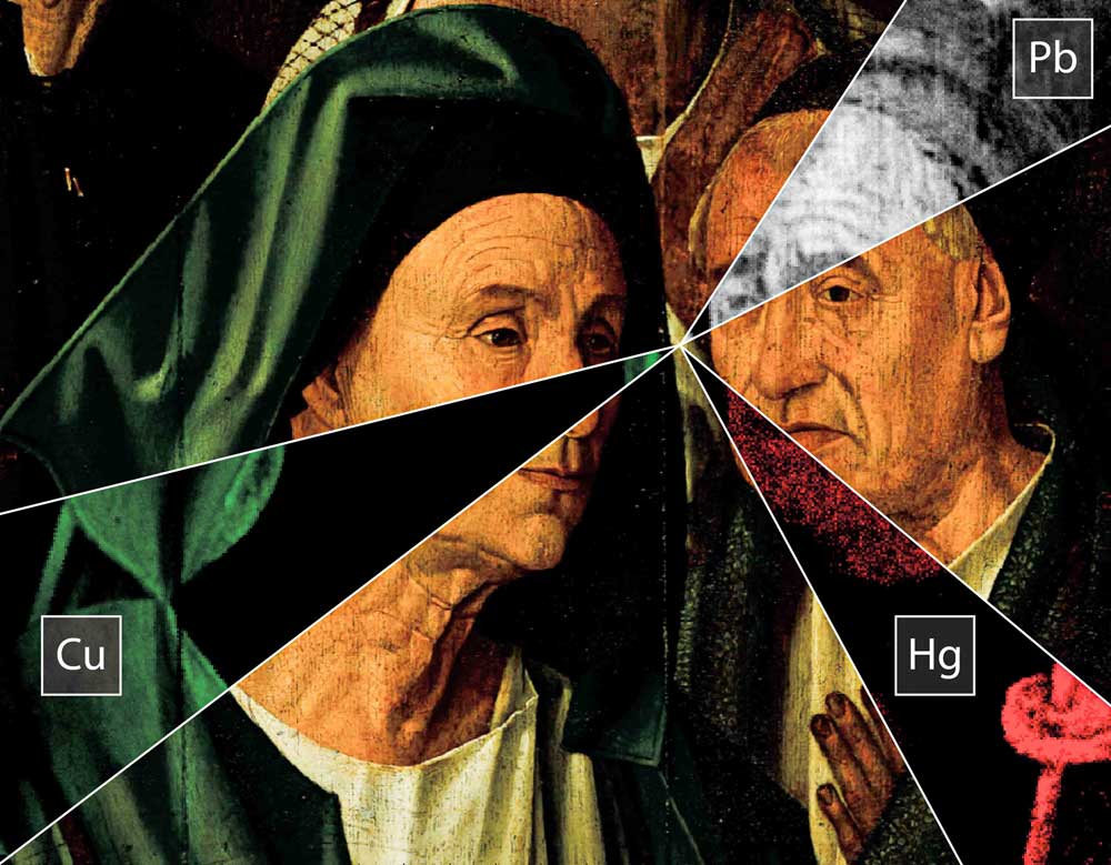
What is "pigment composition?This pigment is typical of the painter or age?Whether in painting hidden another layer of paint?"
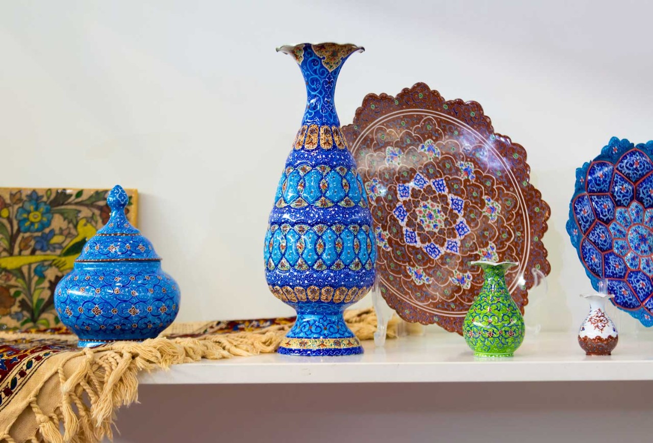
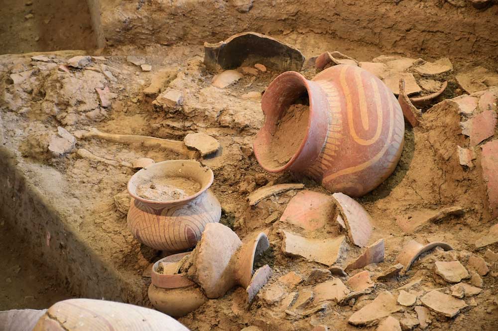
Much attention has been paid to these problems in the aspect of art archaeology and identification.But in principle is not limited to the painting.In the research on the source of all kinds of cultural relics, XRF is a key technology.
ELIO is used to measure the distribution and element can avoid the valuable works of art such as painting or historical relics.If cultural relics is too heavy or too large, the transport may not be feasible, such as rock and cave paintings of testing.Brooke ELIO is the only truly portable XRF (PXRF) imaging spectrometer.Open design system design allows the samples at the scene to non-destructive analysis.
Brooke ELIO option can be used to measure or use of imaging in the regions of 100 mm x 100 mm field distribution of the detection of multiple elements at the same time.In such a large area to collect XRF spectrum is also referred to as macro - XRF or MA - XRF.
application

Technical parameters
The technical details
Motivate and filter options |
|
Analysis of the range |
|
The detector |
|
scanning |
|
collimator |
|
Size and weight |
|
webinar


News and events
For more information
Resources and publications
Brochures and leaflets
Published an article
- 2020 - Journal of Cultural Heritage: Pigments and glassy matrix of the 17 th - 18 th century enamelled French watches: A non - invasive on - site Raman and pXRF study
- 2020 - Microchemical Journal: Chemistry of modern paint media: The strained and collapsed painting by Alexis Harding
- 2019 - Archaeological and Anthropological Sciences: First the in situ pXRF analyses of rock paintings in Erongo, Namibia: the results of the current limits, and prospects
- 2019 - Mesoamerican Manuscripts: New Scientific Approaches and Interpretations: he Early Americas: the History and Culture
- 2019 - Spectrochimica Acta Part A: 雷竞技怎么下载Molecular and Biomolecular Spectroscopy: The In - situ technical study of modern paintings Part 1: The evolution雷竞技网页版 of artistic materials and painting techniques In ten paintings from 1889 to 1940 by Alessandro Milesi (1856-1945),
- 2019 - Journal of Anthropological Archaeology: Social and technological changes in the ceramic production of the Northern Levant during the LBA/IA the transition: New evidence about the Sea People issue through archaeometry
- 2018 - the Heritage Science (Open Access) : Multianalytical approach for the analysis of the Codices Millenarius Maior and Millenarius Minor in Kremsmuenster Abbey, Upper Austria
- 2016 - the Heritage Science (Open Access) : Disclosing Jackson Pollock 's the palette in Alchemy (1947) by non - invasive spectroscopies
- 2016 - Meteoritics and Planetary Science (Open Access) : The meteoritic origin of Tutankhamun 's iron dagger blade
- 2016 - the Heritage Science (Open Access) : the study of the an enameled glass mosque lamp: a 13 th - 14 th - century Mamluk example or 19 th - century European version?