SKYSCAN 1272

Bright spot
Bright spot
SKYSCAN 1272 - micro - high resolution computed tomography (CT)
Based on computed tomography (CT) scanning microscopic imaging technology (micro - CT) SKYSCAN CMOS 1272, is an innovative, high resolution desktop 3 d X-ray microscopic imaging system, integrates the latest X-ray technology.
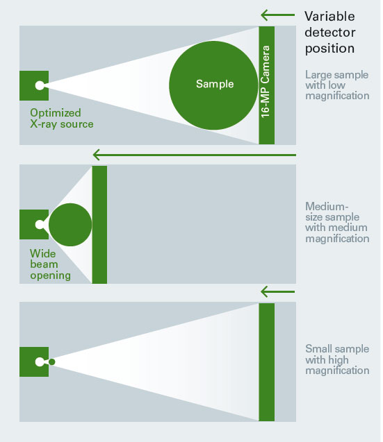
Industry-leading 16 million pixels sCMOS X-ray detector provides excellent resolution, can generate high contrast images.Extension of probe field of vision and enhanced X-ray sensitivity shorten the scan time by one times.Each section of the original resolution up to 11200 x 11200 pixels, can without rescanning samples down to any part of the 3 d volume.New Clean Image ™ scanning mode from the start, greatly reducing the typical CT artifacts, which provides the high quality Image, without tedious after correction.
Top performance with very low cost of ownership.Brooke desktop SKYSCAN 1272 CMOS version can be placed in any laboratory experiment platform, do not need to take up a lot of expensive laboratory space.Just a standard household power plug can run, also does not require water or additional compressor.In addition, the seal of industrial X-ray source is free of maintenance, and therefore there is no other hidden costs.SKYSCAN 1272 carrying 3 d. SUITE software.This comprehensive package covers the GPU acceleration reconstruction, 2 d / 3 d morphology analysis and visualization of surface and volume rendering.
The characteristics of
The main features
Genius mode
SKYSCAN 1272 with Genius mode can be automatically selected parameters.Just click once, then automatically optimize power, energy, filtering, exposure time and background correction.
Moreover, the large size can make samples and sCMOS camera as much as possible close to the light source, it can significantly increase the measured signal intensity.It is for this reason, SKYSCAN 1272 scanning speed than the conventional system detector position fixed up to 5 times faster.
Automatic sampler
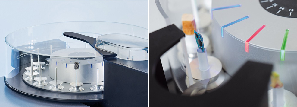
SKYSCAN 1272 can choose to cooperate with a 16 position of external automatic sampler, to quality control and routine analysis in order to increase processing speed.
Automatic sampler can accommodate different sizes of samples, the samples up to 25 mm in diameter.
Can easily switch samples at any time, not break the ongoing scanning process.System can automatically detect the new sample, the LED can display each scanning state: preparation, scanning and complete.
In situ test rig
Brooke's material test bench can be maximum compression test of 4400 N and 440 N tensile test.All test bench can be contacted automatically by the system of the turntable, without any external cables.Through the use of software provided, can set the scheduled scan test.
Brooke's heating and cooling units can reach the highest + 80 DHS C or below the lowest environment temperature low temperature of 30 DHS C.And other test bench, heating and cooling units do not need any extra connection, the system can automatically identify the different test rig.Through the use of heating and cooling, the test samples under the condition of the environment, to assess the micro structure of the temperature of the sample.
application
The key application
Bone application

SKYSCAN 1272 lasted for 1072 and 1172 SKYSCAN SKYSCAN, their contribution to the whole of the micro - at least half of the CT bone shape measurement of literature.It is the benchmark of high resolution, high flux, and ease of use.This makes it a bone disease model (from osteoporosis and osteoarthritis to bone cancer and multiple myeloma) and large-scale genetic phenotypes of ideal solution.
SKYSCAN benchtop instruments have the ultimate performance of voxel size is 0.4 um, 20-100 kV X-ray voltage, provides the resolution and contrast to analyze tube bone bone fracture and blood vessels.
High throughput and automatic batch scanning, including an optional sample changer, 16 samples can be loaded in advance, this is a powerful set of tens of thousands of phenotypic, genetic screening or zebrafish trabecular and cortical bone parameters in mice.
Advanced 3 d image analysis function including phase contrast/retrieval (Paganin, small Angle), 3 d registration, can solve all the flexible model and analysis requirements.
Bone shape measurement (ASBMR nomenclature), has a full 3 d and 2 d parameters of density measurement includes preclinical size within the scope of the BMD of the calibration reference.
Soft tissue

- The typical application of this system is a visualization of mouse lung tumor growth or blood vessels.Emphysema and fibrosis is clearly visible.
- The ultimate performance of the desktop model, voxel size is 0.4 um, 20-100 kV X-ray voltage, provide the resolution and contrast to alveolar level.
- Stability in the process of scanning these fragile or wet tissue can be a challenge, the company developed a special set of brooke specimen can solve this problem.
- Complete internal software solutions for morphological analysis, lung tumor in CT - Analyser software automatic separation of the lungs and other tissues.
Plants and animals

Micro - CT for plant and animal tissues the most remote part of the internal structure of the visual effect is very good.This imaging method can be without damage or destruction of scanning object density.This is why the zoology and botany is the rapid development of micro - CT applications, while SKYSCAN 1272 users walk in the forefront.Can use or at least do not need to sample processing, visualization and analysis of a variety of different organisms.
- High resolution scanning, the object detection as low as 0.4 um, combined with 11 million or 16 million megapixel camera, the useful volume to achieve high resolution.
- Due to the continuous voltage range of the source and 6 internal energy filters, can provide perfect scan for each sample set.
- Comprehensive ability of 3 d image analysis, including the shape and density measurement, phase contrast/retrieval (Paganin, small Angle), 3 d registration, segmentation and advanced image processing methods.
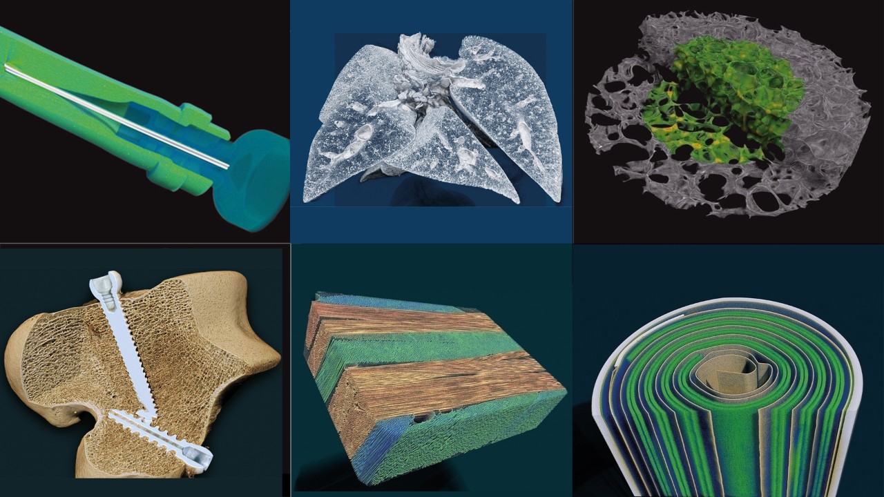
Application scenarios
The profile of kidney stones, in order to 2 microns voxel size scan, according to different stages of development.(sample by Belgian Sart Tilman liege university hospital clinical chemistry lab)

3 d model of the femur in mice with 5 micron voxel size scanning, the local thickness of color coding.

Cross-sectional tibia by mice, to 800 nanometers voxel scan, showed bone cells in lacunae.

3 d renderings of tibia in mice to 800 nanometers voxel scan, showed bone cells (blue) and vascular network (red).

Through the mice lungs cross-sectional to 1 micron voxel size.

To 10 microns voxel size scan stag beetles.Provide the sample of the university of Antwerp, Belgium.

With 5 micron voxel size scanning of coffee beans.

The internal structure of the 3 d rendering rose.

3 d rendering of polylactic acid scaffold pore network of color coding, local thickness of 1.5 microns voxel size scan.

To 875 nm voxel size scan wood sample cross section.

At 8.9 microns voxel size scan sand crabs 3 d rendering.

Pixel size 17 microns to scan the sea urchin shell.

Mouse embryos (E16.5) orthogonal cross section to 1.75 micron voxel size scan.

Volume rendering 3 d model of mice with pulmonary vascular and trachea, chemical drying to 10 microns voxel size after scanning.In the center of the left lung a necrotic area can be observed.

Bees head 3 d models: PTA high resolution scanning after dyeing.The right of the model is a virtual cutting, to reveal the internal structure.From the university of granada of (sample)

Technical parameters
SKYSCAN 1272 CMOS technology parameters
The characteristics of |
parameter |
advantage |
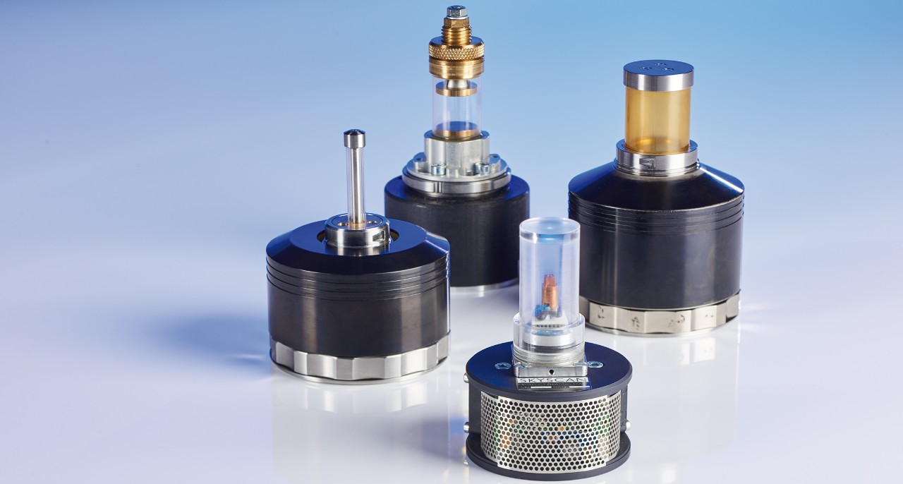
X-ray source |
40-100 kV 10 W. < 5 m (including size, 4 W |
Maintenance free, fully sealed X-ray source Quality control was achieved by rapid scanning, or 4 d XRM |
X-ray detector |
16 MP sCMOS detector (4096 x 4096 pixels) | Precision of the detector can achieve the highest resolution |
The sample size |
The biggest diameter of 75 mm Maximum height of 70 mm |
Suitable for small to medium size samples |
Automatic sampler (optional) |
16 kinds of sample, up to 25 mm in diameter The outer |
Unattended, high flux The size of the sample random combination Add/remove samples at any time, don't interrupt the scanning process |
size |
1160 mm x 520 mm x 330 mm (deep x width x height) Weight 150 kg |
Save a space, suitable for every lab desktop system |
The power supply |
100-240 - v AC, 50 to 60 hz, the biggest 3 a | Minimum standard installation requirements, the power supply can meet the requirements |
software
Orientation, scanning, reconstruction and analysis

Brooke XRM solution contains all the software needed to collect and analyze data.Intuitive graphical user interface combined with the user guide of parameter optimization, suitable for both professional users and is suitable for novice users.By using the latest GPU acceleration algorithm, reconstruction time is greatly shortened.CTVOX, CTAN and CTVOL combined, forming a powerful software suite, support for qualitative and quantitative analysis model.
Measuring software:
SKYSCAN 1272, instrument control, measure planning and collection
Reconstruction of software:
NRECON - 2 d figure projection drawing into a 3 d volume
Analysis software:
DATAVIEWER - check the 3 d volume step by step, the 2 d / 3 d image registration
CTVOX - through body rendering shows a real situation
CTAN - 2 d / 3 d image analysis and processing
CTVOL - surface model visualization, can be exported to a CAD or 3 d printing
For more information
SKYSCAN 1272 related video
Easily scan 16 samples
Start your analysis!
What is the XRM?
support
Service and support
We offer:
- Professional senior technical personnel to do fault professional technical support, isolation and solve the problem of hardware and software
- Network remote service, providing diagnosis and application support
- Fusion reality support - virtual engineers in your side,video)
- According to your request to schedule maintenance
- Customer site maintenance, maintenance services
- Spare parts delivery will be within one working day/the world needs a few working days to complete
- Installation qualification and operational qualification/performance verification of compliance services
- Site planning and relocation
- Find nextA training course
Check out ourSupport site:
- Software updates
- Product manuals and installation guide
- Training video
Need to register.
LabScape
Brooke BioSpin nuclear magnetic resonance (NMR) and preclinical imaging products, services and lifecycle support
Brooke promises to provide clients with unparalleled throughout the buying cycle, from the initial consultation to the evaluation, installation, and service life of the instrument, it is LabScape always adhere to the service concept.
LabScape Maintenance agreement (Maintenance Agreements), the optional services (On - site On - Demand) and laboratory improvement plan (Enhance Your Lab) modern Lab is dedicated to provide you with a new method of Maintenance and service.