NanoWizard V BioScience
Destaques
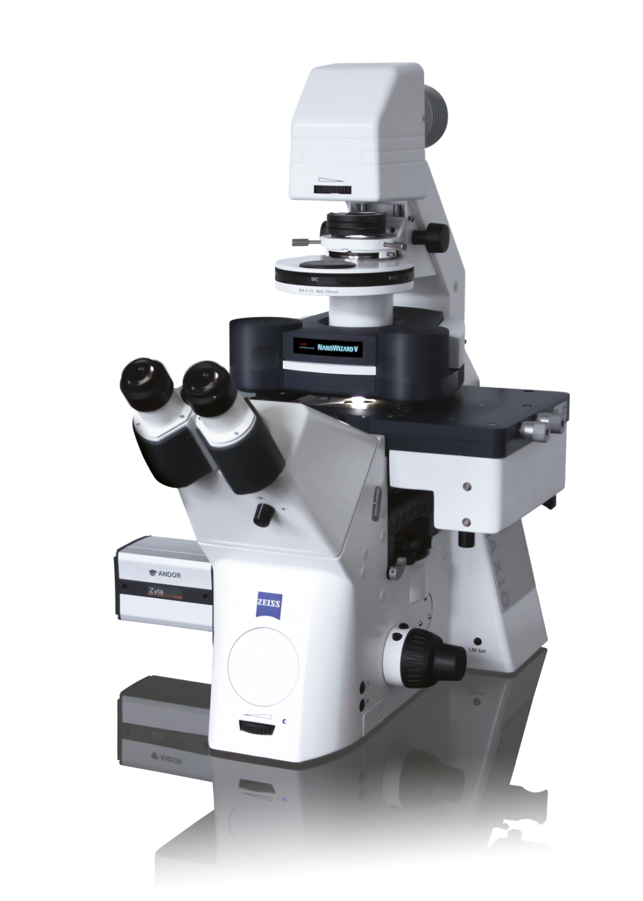
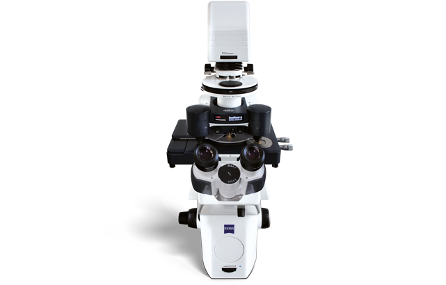
NanoWizard® V BioScience
The JPK NanoWizard® V combines high spatio-temporal resolution with a large scan area, flexible experiment design, and out-standing integration with advanced optical microscope systems. The automated setup, alignment, and re-adjustment of system parameters opens new possibilities for long-term, self-regulating experiment series.
Características
Discover the 5th Generation BioAFM
NanoWizard Vis expected to significantly advance our understanding of dynamic cellular processes and molecular mechanisms. Its PeakForce-QI mode enables fast and flexible quantitative nanomechanical measurements—significantly extending the capabilities of AFM—while its automated, remote-control, and fast-scanning capabilities provide high-throughput, high-performance imaging of even complex experiments.
NanoWizard Vfeatures novel scanner and sensor technologies and state-of-the-art control software that encompasses an intuitive, workflow-based graphical user interface (GUI) to ensure true, easy-to-use AFM operation.
- Unmatched ease of use
- Speed for dynamics and improved throughput
- Automated, high pixel density mapping and imaging
- From the pioneers of BioAFM withover 25 years of experiencein developing BioAFMs
- Recognized by an install base ofover 1000 JPK/Bruker BioAFMsacross the globe
- Provenby 8500+ publicationswith biological significance
- Supported by dedicated cantilever development for high-resolution imaging and customized applications
- 巴勒斯坦权力机构ving the way for new scientific discoveries with:
· PeakForce-QI, PeakForce Tapping®, PeakForce QNM®, QI
· Single Molecule Force Spectroscopy (SMFS)
· Single Cell Force Spectroscopy (SCFS)
· DirectOverlay 2 for AFM in conjunction with advanced optical microscopy
· New software environment V8
· Features the latest ExperimentPlanner and ExperimentControl
· Accessories for high NA optics and AFM, environment control, and more
Latest Technology for Maximum Efficiency
Highest performance BioAFM
Featuring an enhanced, workflow-based design with intuitive user guidance in the new SPM V8 software, the NanoWizard V empowers beginners and experts alike to acquire highest quality, reproducible data. The system’s high degree of automa-tion improves productivity and maximizes throughput. State-of-the-art analysis and batch processing routines ensure scientific accuracy and statistical data reliability.
- Adaptive intelligent scanning routines enable faster scanning rates of up to 400 lines/sec
- Lowest noise scanner and detection system ensure high-resolution data and unrivalled performance on inverted optical microscopes
- PeakForce-QI, the symbiosis of PeakForce Tapping and QI modes, delivers the fastest, most advanced force control for highly delicate samples
Fastest automated BioAFM for corrugated samples on an inverted microscope
- Visualize dynamics with Bruker’s Nested Scanner and latest feedback technologies
- Bruker’s proven DirectDrive increases cantilever excitation stability
- Active balancing provides faster scanning over large scan ranges
- Improved productivity and maximized throughput enables better statistics
- Intelligent optimization routines deliver quantitative nanomechanics
Most intuitive BioAFM operations
- Fundamental ease-of-use features
- User management, ideal for multi-user facilities
- Automated setup and workflow
- Single-click cantilever calibration
- Single-click optical image calibration
- Extended optical viewing field for AFM with stitching feature
- Long-term, unattended experimental procedures
- Remote operation capabilities
- Optimized storage of parameters and favorites
- Intuitive integration of data processing routines
"The NanoWizard V is excellent for medical component analysis."
Prof. Dr. rer. nat. Hans Bäumler
Head of Research Department
Institute of Transfusion Medicine, Charité University Hospital Berlin (Germany)

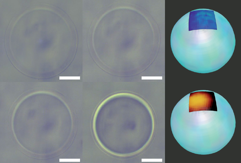
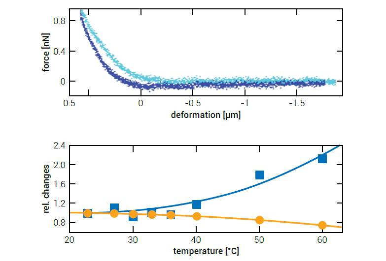
Understanding Forces in Biology
The leading BioAFM for mechanobiology
By providing the right model for the right experiment, Bruker delivers easy solutions to complex scientific questions. Automated measurement procedures allow researchers to concentrate on what’s important - their research - and to question why and not how.
- Investigate single molecules, live cells, and tissue samples
- Study corrugated and delicate, soft sample structures under native conditions
- Gain a detailed understanding of viscoelastic properties and adhesion processes
- Highest level of automation thanks to Autoalign, HybridStage or motorized stage, ExperimentPlanner, ExperimentControl, and GUI featuresfor high-speed signal processing and lowest noise levels
PeakForce-QI – a new chapter in quantitative imaging
The pioneers of quantitative biological imaging, with PeakForce Tapping (2000+ publications) and QI (~1000 publications), now bring you the next generation:
PeakForce-QI.
- Fastest nanomechanical imaging thanks to latest piezo and sensor technologies
- Easy, precise batch processing
- True, real-time force curve monitoring
"The system’s promised speed and resolution, ease of use, and up-to-millimeter-range capabilities make this a game changer for AFM investigations in nanomedicine and bio-medical applications."
Dr. David Martinez Martin
Physicist · Senior Lecturer in Biomedical Engineering
Co-chair of the sensors and diagnostics cluster of the Nanohealth Network (University of Sydney)

Quantitative Data and Intelligent Analysis
Automated Force Spectroscopy Reloaded
The pioneers of Single-Molecule Force Spectroscopy (SMFS) and Single-Cell Force Spectroscopy (SCFS) now bring you even more flexibility, higher precision, and increased throughput.
- Proven ForceRobot technology now built-in
- Bruker’s intuitive and powerful RampDesigner
- Automated calibration
- Molecular recognition imaging
- Most sensitive force control and tip-saving features
Powerful Data-Slices
- Flexible creation of topography images at different forces
- Proprietary contact point imaging
- 图像叠加输出的通道从批处理过程ssing
For complex experiments, from single molecules to cells and tissues
- Optimized environmental control options
- Comprehensive nano-mechanics with RampDesigner
- Complex experimental routines with ExperimentPlanner
- Integration of several modes in long-term, unattended experiments
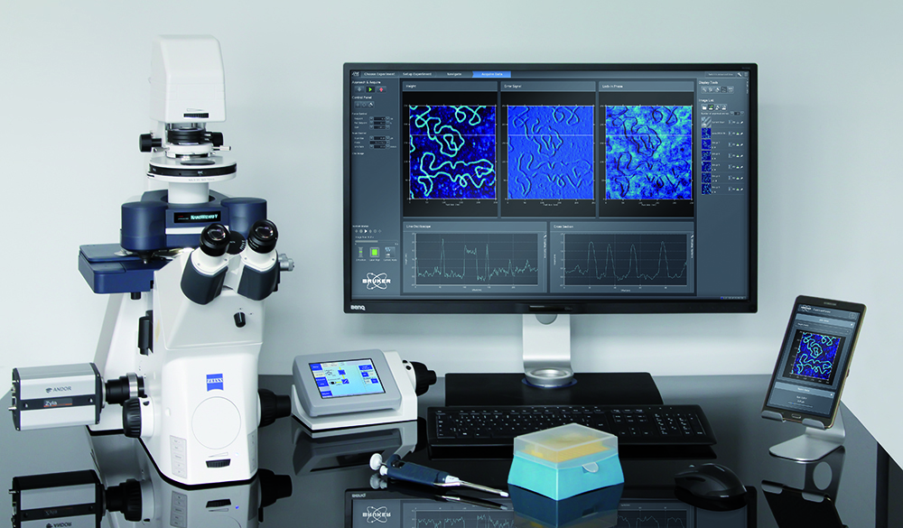
Correlated Microscopy Newly Defined
Leading BioAFM in combination with advanced optical microscopy
- Perfect integration into advanced optical techniques
- Optical super-resolution (STED, FLIM, …)
- Upright for tissues, implants etc.
- DirectOverlay 2/MIRO
- One-click optical image import
- 980 nm OBD option
- DirectTiling, for larger scale efficiency
- Stitching
- MultiScan
- Seamless integration of multiple optical, external and AFM channels
Optical Modes Combinations
- Brightfield
- DIC
- Phase or modulation contrast
- Confocal microscopy
- Spinning disc
- TIRF and IRM
- FRET, FLIM, FRAP, FCS
- Ca2+imaging
- Superresolution (STED, PALM/STORM, SIM)
- Macroscope combination
- Optical tweezers with OT-AFM
Unrivalled Flexibility by Design
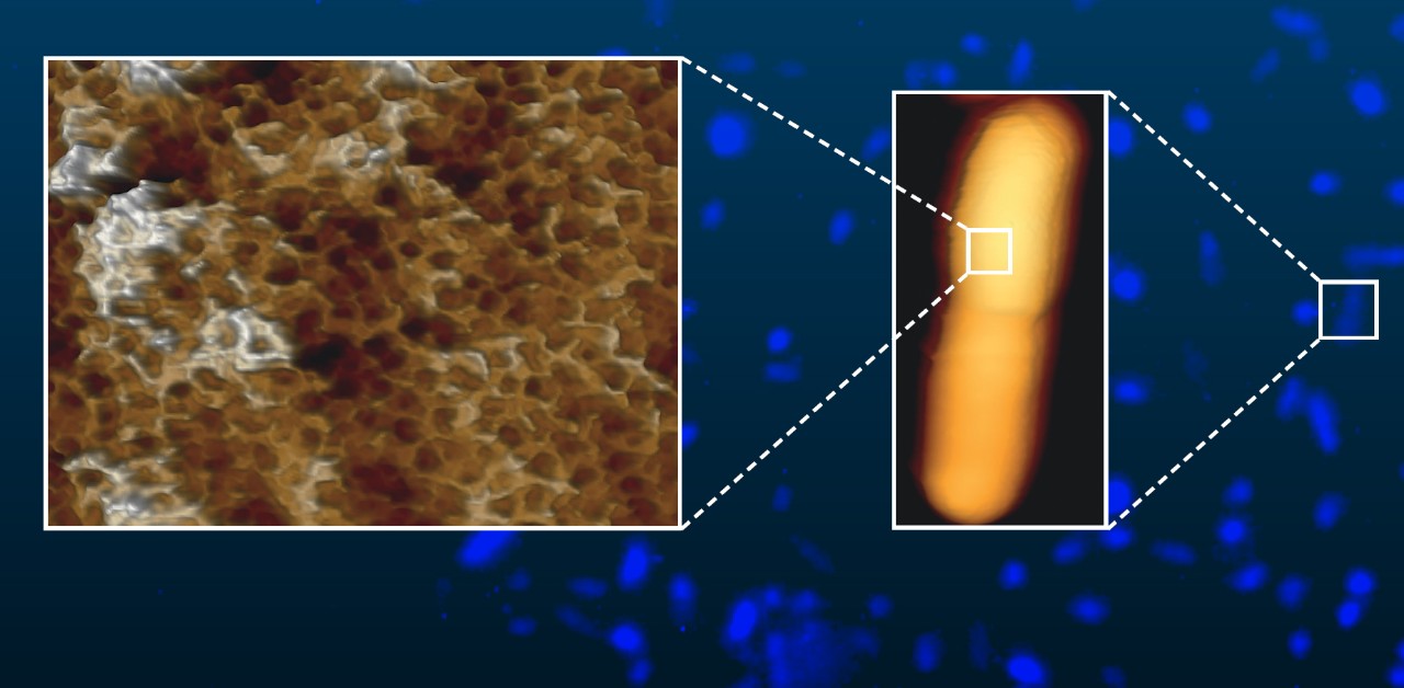
Optimized for use in microbiology and virus research
The NanoWizard V BioAFM enables direct access to pathogens and infectious agents in a BSL facility. Each step, from sample preparation, loading and disposal to running the experiment, can be performed directly in the biosafety lab.
- Suitable for working with native pathogens and biohazardous material
- No “additional preparation“ steps
- BSL-3 compliance achieved
(in accordance with lab’s safety regulations)
Explore an extensive range ofaccessoriesfor versatile experimental setups and environmental control.
Seamless combination with theFluidFM technologyenables a host of novel experiment designs and applications.
Aplicações
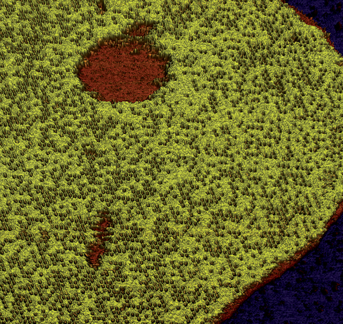
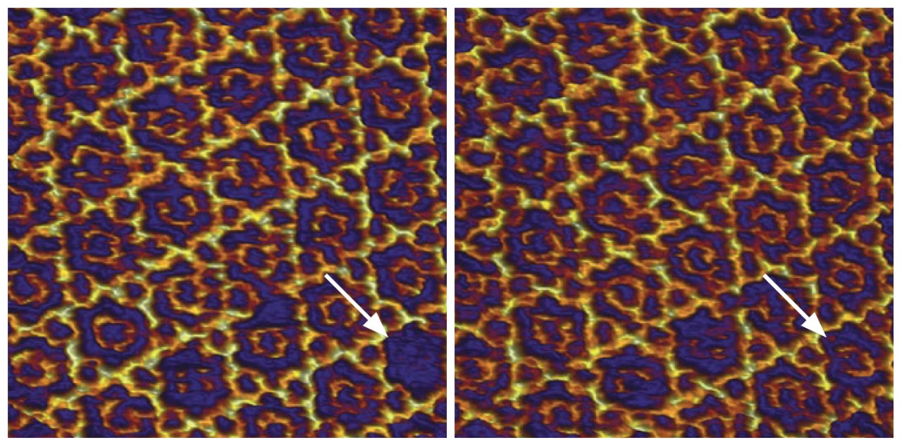
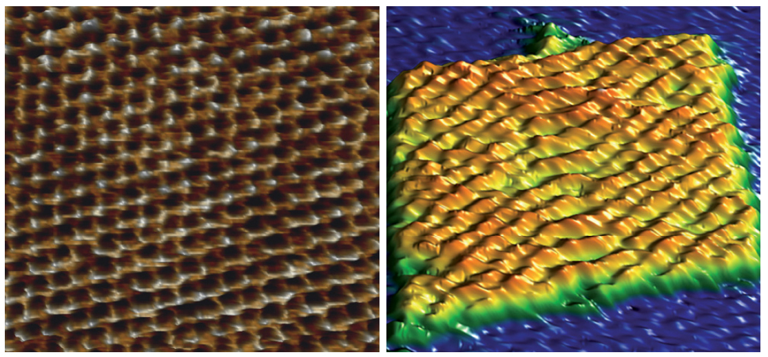
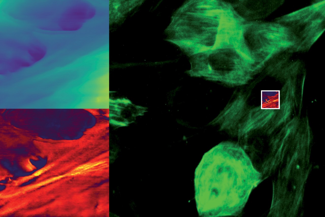
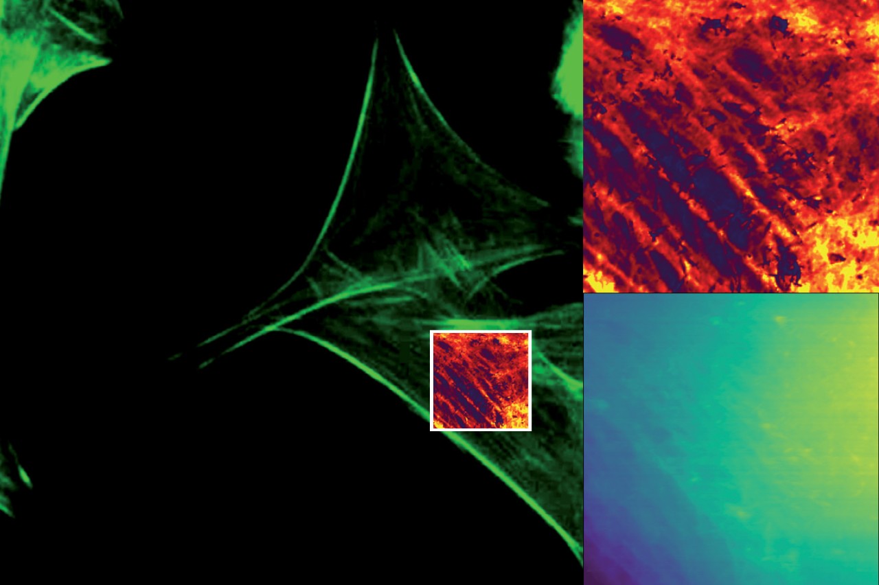
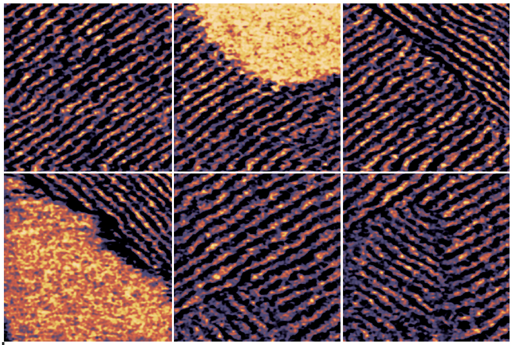
Life Science AFM Data Gallery
Bruker’s BioAFMs allow life science and biophysics researchers to further their investigations in the fields of cell mechanics and adhesion, mechanobiology, cell-cell and cell-surface interactions, cell dynamics, and cell morphology. We have collected a gallery of images demonstrating a few of these applications.
Especificações
Operating Modes
Standard Operating Modes
- Now with PeakForce-QIincluding PeakForce Tapping, QI and PeakForce QNM
- Including fast PeakForce Tapping and QI withnested scannertechnology
- Contact mode with lateral force microscopy (LFM)
- Tapping Mode™ with PhaseImaging™
- ExperimentPlannerfor designing a specific measurement workflow
- Static and dynamic force spectroscopy
- Advanced Force Mapping
Optional Modes
- Advanced spectroscopy modes such as various force clamp modes or ramp designs
- Fast scanning optionwith line rates of up to 200 Hz
- QI Advanced mode for quantitative data, perfect for soft samples
- ScanAsystautomated gain and setpoint adjustment in PeakForce Tapping and PeakForce-QI
- Advanced AC modes such as FM and PM with Q-control & Active Gain Control
- Microrheology inCellMech Package
- Kelvin Probe Microscopy
- MFM and EFM
- Conductive AFM
- STM
- Electrical spectroscopy modes
- Piezoresponse Microscopy for high voltages
- Electrochemistry & Scanning Electrochemistrywith temperature control and optical microscopy
- NanoLithography and NanoManipulation
- NanoIndentation
- Scanning Thermal AFM
- FluidFM®solution from Cytosurge
- ExperimentControl feature for remote experiment control
- DirectOverlay 2 for combined AFM and optical microscopy
- Additional XY or Z sample movement stages available withCellHesion®, TAO™ and HybridStage™ module
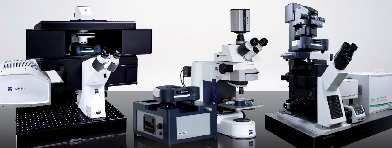
Acessórios
The Widest Range of Accessories in the Market
Optical systems/accessories, electrochemistry solutions, electrical sample characterization, environmental control options, software modules, temperature control, acoustic and vibration isolation solutions and more. Bruker provides you with the right accessories to control your sample conditions and to perform successful experiments.
Webinars
Watch Recent BioAFM Webinars
Our webinars cover best practices, introduce new products, provide quick solutions to tricky questions, and offer ideas for new applications, modes, or techniques.